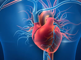
Angio is a life-saving examination method used in the diagnosis of cardiovascular diseases in sudden heart attacks. It shows the problem in the vessels going to the heart and treatment planning is made according to the result.
Coronary angiography is usually performed through the artery (A. femoralis) located in the patient's right inguinal region. If this vein is not used (for example, this vein may be completely occluded), the left inguinal artery, right-left wrist arteries, elbow or axilla arteries can also be used. The procedure takes about 10-20 minutes, and this time may take 20-30 minutes in patients with by-pass. Before the procedure, the area to be angio is anesthetized with local anesthesia and the patient does not feel any pain. After the procedure is over, the cannula placed on the intervention site is withdrawn, pressure is applied for 15-20 minutes to stop the bleeding, at the end of the 6th hour, the patient is stood up and discharged. You can take a bath the next day and return to normal life if there is no swelling, uncomfortable pain or extensive bruising at the site of the intervention.
Are there risks to angiography?
Although coronary angiography is a procedure that is mostly performed without any problems, it also carries a risk of complications since it is an interventional examination method. Angio risks can be described as follows: The risk of death, defined as major complications, is 1 in 1400 cases (0.07%); stroke 1 in 1000 (0.1%); coronary artery injury 1 in 1000 (0.1%); arterial access site complication is 1 in 500 (0.2%). The more common minor (minor) complications are temporary and are bleeding or hematoma (blood clot) at the entry site, false ballooning, rhythm disturbances, sensitivity to the dyed substance used, and vagal reaction (nausea, vomiting, cold sweat, low blood pressure, pulse rate). slowing of the cannula usually occurs during insertion or withdrawal of the cannula). The cardiologist will inform about the complications that occur after angiography.
Balloon Angioplasty Procedure:
Coronary Balloon Angioplasty is the procedure of widening the clogged vessel before the stent, by continuing the procedure in the same session, for patients whose coronary angiography has been decided to apply a balloon to the diseased vessel. Strictures are removed by controlled inflation of the specially designed balloon in the stenosis area within the vessel. The procedure usually takes less than half an hour and the patient who does not need long-term medication is usually discharged the next day.
Coronary Stent rare?
Coronary stents have been developed to provide a better blood flow in the vessel where full patency is not provided in balloon treatment and has been widely used since the 90s. Coronary Stent (steel wire cage) is a method used to eliminate these problems in patients with intravascular rupture after the balloon procedure. stent; It is placed on the balloon and when the balloon is inflated in the vessel, it is mounted on the inner wall of the vessel. The success rate in balloon and stent application is between 65-99%. Re-narrowing (restenosis) may occur with a probability of 20-30% within a six-month period. In newly introduced drug-coated stents, this probability has decreased to the range of 8-15%. In case of narrowing in the stent, the balloon or stent can be applied again.
After stent placement, the patient can be taken to the coronary intensive care unit. Hospital stay is usually 24 hours.
Pacemakers
The heart is a complex organ that works with electrical impulses. With these electrical transmissions, regular contraction function, which is the main function of the heart, is provided. With contraction, the blood needed by the body is pumped and our oxygen needs are met. However, some problems that occur in the formation of signals or in the electrical transmission system that provides the transmission of the signals cause the deterioration of this perfect system. The contraction pattern of the heart is disturbed, the heart weakens and serious health problems may occur. Pacemakers are used in the treatment of problems that occur in the heart's stimulus system or the electrical transmission system that transmits the stimulus. There are different types of pacemakers specific to different diseases. Thanks to the shock treatment given by pacemakers and pacemakers with shock properties, the lifespan of patients is prolonged and pacemakers play a life-saving role in the treatment of many serious problems.
Rhythm Disorder and Ablation
The heart, which works regularly between 60 and 100 per minute, experiences a rhythm disorder (arrhythmia) as a result of the deterioration of this harmony. .. While this arrhythmia is occurring, the heart may beat too fast (tachycardia), too slow (bradycardia), or irregularly. And the pump function of the heart is impaired. Although most arrhythmias are harmless, some of them can be serious enough to cause life-threatening. For example, during arrhythmia, the heart cannot send enough blood to the body, and this can cause shortness of breath, fainting, fainting, and even sudden death.
The abnormal heart tissue that causes tachycardia is destroyed and removed, so the discomfort can be treated. This treatment is called catheter cardiac ablation, radiofrequency ablation (RF ablation), cardiac ablation or simply ablation. The catheter ablation procedure is similar to EPS or coronary angiography. The patient is first taken to the catheter laboratory. Generally, local anesthesia is applied to the inguinal region of the patient and it is entered through the inguinal vein. It is a painless procedure. After the responsible area is detected, energy is given to this area by means of a catheter with a metal electrode at the tip, and the responsible cells in this area are burned and destroyed. During this time, the patient may feel burning and palpitations in the chest. The processing time is usually very variable, and the process can take from 1.5 to 6 hours.